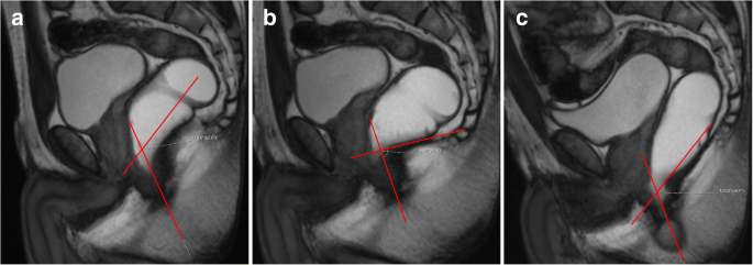
Weakening of the female pelvic floor is a prevalent and debilitating disorder.
Pelvic floor descent radiology.
There are two components of pelvic floor relaxation.
Symptoms such as obstructive defecation incontinence and sphincter complex disorders have a significant impact on patient lifestyle and physical mental well being 1 2.
Pelvic floor descent or descending perineal syndrome occurs when pelvic muscles lose tone resulting in excessive descent of the entire pelvis floor at rest or during evacuation.
Mr imaging clearly shows lack of descent of the pelvic floor during defecation and paradoxical contraction of the puborectalis muscle with failure of the anorectal angle to open thus resulting in prolonged or incomplete evacuation.
Two hundred patients with clinical symptoms suggestive for pelvic floor descent referred to our radiology department for pelvic dynamic mri for the evaluation of pelvic floor disorders.
Imaging studies include colonic transit to assess bowel motility.
Although this condition predominantly affects females up to 16 of males suffer as well.
Damage to other supporting structures within the pelvic floor can only be inferred from the presence of secondary signs and abnormal descent of the pelvic organs during a dynamic mr imaging study.
Symptoms include pelvic pain pressure pain during sex incontinence incomplete emptying of feces and visible organ protrusion.
One anteroposterior with a widening of the puborectal hiatus and one vertical with pelvic floor descent.
On the image subjectively assessed as showing the greatest degree of pelvic.
Pelvic floor dysfunction is an umbrella term for a variety of disorders that occur when pelvic floor muscles and ligaments are impaired.
Pelvic floor imaging is an important part of both gastrointestinal and functional urology urogynaecological departments.
37 2 2 prolapse pelvic organ prolapse pop also called urogenital prolapse is downward descent of the pelvic organs that results in a protrusion through the urogenital hiatus.
It is defined as a line that joins the inferior border of the symphysis pubis to the final coccygeal joint and it is drawn in a midline sagittal image.
Fifty patients satisfied the physical prerequisite to be examined in sitting position and they were asked to take part in the study.
The pubococcygeal line pcl is a reference line for the pelvic floor on imaging studies and helps detect and grade pelvic floor prolapse in defecography studies.
Pelvic floor dysfunction can also be caused by atrophy previous injury to or other weaknesses of levator ani which can lead due global descent of the pelvic viscera due to loss of muscular support.
On dynamic imaging pelvic floor descent is defined as anorectal junction descent of more than 2 5 cm below the pcl.

